Copyright
©2014 Baishideng Publishing Group Inc.
World J Radiol. May 28, 2014; 6(5): 210-217
Published online May 28, 2014. doi: 10.4329/wjr.v6.i5.210
Published online May 28, 2014. doi: 10.4329/wjr.v6.i5.210
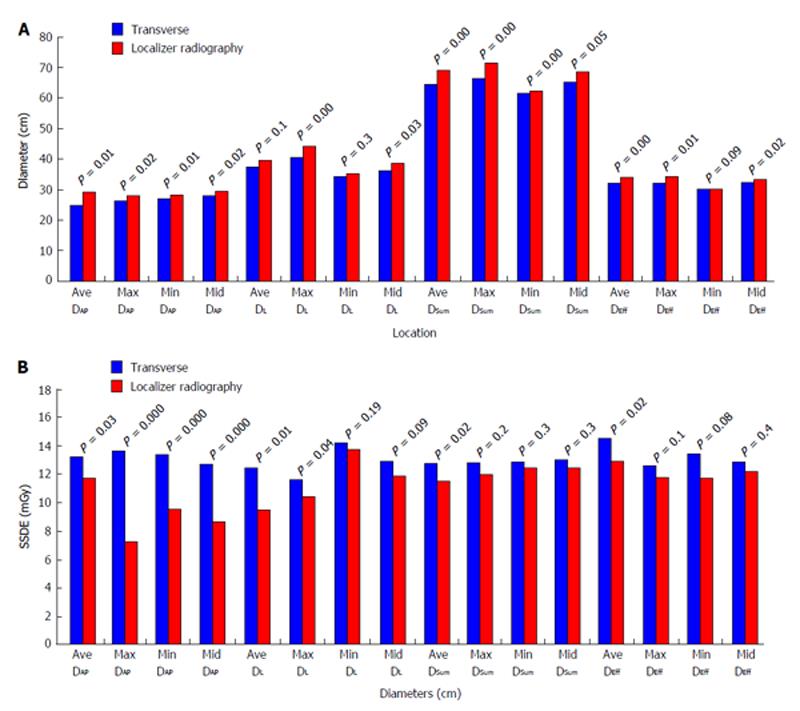
Figure 2 Difference between measurements on localizer and transverse computed tomography images for all values, including averages of interval measurements, maximum, minimum, and mid-scan location are illustrated.
A: Several diameters were measured at different locations from transverse CT images (blue) and localizer radiographs (red). Diameter measurements were significantly different from localizer radiograph as compared to transverse CT images; B: SSDE values obtained from several diameters at different locations from transverse CT images (blue) and localizer radiographs (red). SSDE values were significantly different in maximum and minimum Antero-Posterior diameters. Ave: Average; max: Maximum; min: Minimum; mid: Mid location; CT: Computed tomography; SSDE: Size specific dose estimate; DEff: Effective Diameter; DSum: Sum of anterior-posterior + Lateral diameter; DL: Lateral diameter; DAP:Anteroposterior Diameter.
- Citation: Pourjabbar S, Singh S, Padole A, Saini A, Blake MA, Kalra MK. Size-specific dose estimates: Localizer or transverse abdominal computed tomography images? World J Radiol 2014; 6(5): 210-217
- URL: https://www.wjgnet.com/1949-8470/full/v6/i5/210.htm
- DOI: https://dx.doi.org/10.4329/wjr.v6.i5.210


