Copyright
©2014 Baishideng Publishing Group Inc.
World J Radiol. May 28, 2014; 6(5): 192-202
Published online May 28, 2014. doi: 10.4329/wjr.v6.i5.192
Published online May 28, 2014. doi: 10.4329/wjr.v6.i5.192
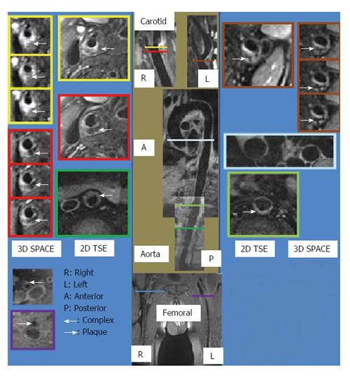
Figure 1 Sample magnetic resonance images obtained from the three vascular beds using both three-dimensional SPACE and two-dimensional turbo spin echo sequences.
Montage showing samples from the two-dimensional (2D) and 3D black blood vessel wall image acquisitions of the carotids, aorta and femoral arteries acquired in a single session (longitudinal sections in the middle, cross sections on each side). Imaging time < 45 min for 3D acquisitions.
-
Citation: Wong SK, Mobolaji-Iawal M, Arama L, Cambe J, Biso S, Alie N, Fayad ZA, Mani V. Atherosclerosis imaging using 3D black blood TSE SPACE
vs 2D TSE. World J Radiol 2014; 6(5): 192-202 - URL: https://www.wjgnet.com/1949-8470/full/v6/i5/192.htm
- DOI: https://dx.doi.org/10.4329/wjr.v6.i5.192


