Copyright
©2014 Baishideng Publishing Group Inc.
World J Radiol. May 28, 2014; 6(5): 177-191
Published online May 28, 2014. doi: 10.4329/wjr.v6.i5.177
Published online May 28, 2014. doi: 10.4329/wjr.v6.i5.177
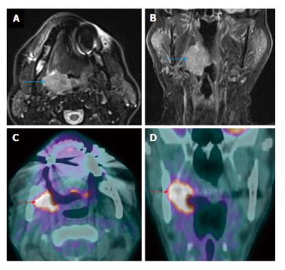
Figure 18 Recurrent primary tumor detected by positron emission tomography/computed tomography.
A T3N0M0 right palatine tonsil squamous cell carcinoma was demonstrated on axial T2W magnetic resonance imaging (MRI) (blue arrow) (A) and coronal contrast-enhanced T1W MRI (B). MRI and positron emission tomography/computed tomography (PET/CT) performed 2 and 3 mo after completion of chemoradiation demonstrated complete treatment response (not shown). Surveilance PET/CT (C and D) revealed intense increase metabolism in the right palatine tonsil and medial pterygoid muscle (red arrows) representing a recurrent squamous cell carcinoma.
- Citation: Tantiwongkosi B, Yu F, Kanard A, Miller FR. Role of 18F-FDG PET/CT in pre and post treatment evaluation in head and neck carcinoma. World J Radiol 2014; 6(5): 177-191
- URL: https://www.wjgnet.com/1949-8470/full/v6/i5/177.htm
- DOI: https://dx.doi.org/10.4329/wjr.v6.i5.177


