Copyright
©2014 Baishideng Publishing Group Inc.
World J Radiol. May 28, 2014; 6(5): 177-191
Published online May 28, 2014. doi: 10.4329/wjr.v6.i5.177
Published online May 28, 2014. doi: 10.4329/wjr.v6.i5.177
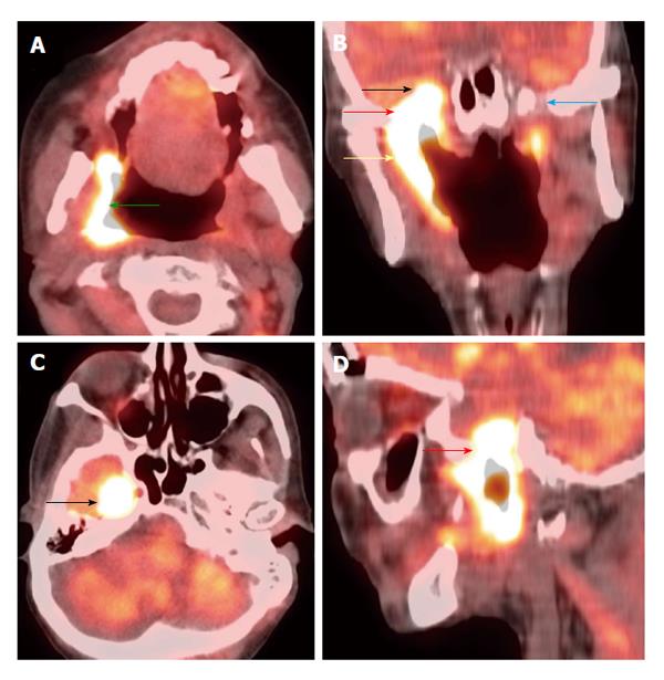
Figure 8 Perineural spread.
Positron emission tomography/computed tomography (A: Axial; B: Coronal; C: Axial; D: Sagittal) demonstrates a T4b right oropharyngeal squamous cell carcinoma (green arrow) spreading along the mandibular branch of the trigeminal nerve (yellow arrow) through the right foramen ovale (red arrows) to involve the right cavernous sinus (black arrows). The left mandibular nerve in the foramen ovale is normal (blue arrow).
- Citation: Tantiwongkosi B, Yu F, Kanard A, Miller FR. Role of 18F-FDG PET/CT in pre and post treatment evaluation in head and neck carcinoma. World J Radiol 2014; 6(5): 177-191
- URL: https://www.wjgnet.com/1949-8470/full/v6/i5/177.htm
- DOI: https://dx.doi.org/10.4329/wjr.v6.i5.177


