Copyright
©2014 Baishideng Publishing Group Inc.
World J Radiol. May 28, 2014; 6(5): 177-191
Published online May 28, 2014. doi: 10.4329/wjr.v6.i5.177
Published online May 28, 2014. doi: 10.4329/wjr.v6.i5.177
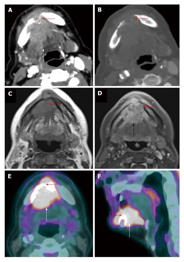
Figure 3 Positron emission tomography/computed tomography for detection of mandibular invasion.
A T4a floor of mouth squamous cell carcinoma (white arrow) with through-and-through involvement (red arrow) of the anterior mandible is demonstrated on axial contrast-enhanced computed tomography (CT) (A: Soft tissue window; B: Bone window), axial contrast-enhanced magnetic resonance imaging (C: T1WI; D: Post contrast enhanced T1WI with fat saturation), and positron emission tomography/CT (E: Axial; F: Sagittal).
- Citation: Tantiwongkosi B, Yu F, Kanard A, Miller FR. Role of 18F-FDG PET/CT in pre and post treatment evaluation in head and neck carcinoma. World J Radiol 2014; 6(5): 177-191
- URL: https://www.wjgnet.com/1949-8470/full/v6/i5/177.htm
- DOI: https://dx.doi.org/10.4329/wjr.v6.i5.177


