Copyright
©2014 Baishideng Publishing Group Inc.
World J Radiol. May 28, 2014; 6(5): 177-191
Published online May 28, 2014. doi: 10.4329/wjr.v6.i5.177
Published online May 28, 2014. doi: 10.4329/wjr.v6.i5.177
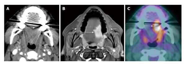
Figure 1 Dental amalgam artifact obscuring an oral cavity tumor.
A: The T2 left lateral oral tongue squamous cell carcinoma (arrow) was obscured by the dental amalgam artifact on contrast-enhanced computed tomography (CT); B: The lesion is better evaluated on the contrast-enhanced T1W; C: Positron emission tomography/CT (PET/CT) images. Information from the PET portion is clearly less affected by streak artifact. However, PET/CT does not add additional information to magnetic resonance imaging in terms of primary oral cavity lesion extent for the majority of cases.
- Citation: Tantiwongkosi B, Yu F, Kanard A, Miller FR. Role of 18F-FDG PET/CT in pre and post treatment evaluation in head and neck carcinoma. World J Radiol 2014; 6(5): 177-191
- URL: https://www.wjgnet.com/1949-8470/full/v6/i5/177.htm
- DOI: https://dx.doi.org/10.4329/wjr.v6.i5.177


