Copyright
©2014 Baishideng Publishing Group Inc.
World J Radiol. May 28, 2014; 6(5): 169-176
Published online May 28, 2014. doi: 10.4329/wjr.v6.i5.169
Published online May 28, 2014. doi: 10.4329/wjr.v6.i5.169
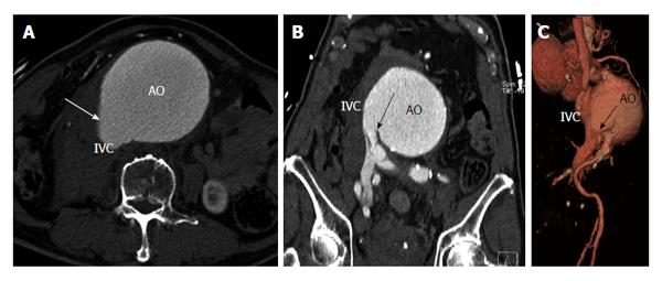
Figure 13 Aorta to inferior vena cava fistula.
A: Axial contrast computed tomography (CT) of the abdomen in a patient with history of abdominal aortic aneurysm (AO) shows similar contrast opacification of the inferior vena cava (IVC). There is a communication demonstrated (arrow) between the aorta and IVC; B: Coronal contrast CT of the abdomen demonstrates the site of fistulous communication between the aorta and the distal IVC adjacent to the right common iliac vein; C: Volume rendered 3D computed tomography angiography shows fistulous communication (arrow) between the abdominal aorta and IVC.
- Citation: Ghandour A, Rajiah P. Unusual fistulas and connections in the cardiovascular system: A pictorial review. World J Radiol 2014; 6(5): 169-176
- URL: https://www.wjgnet.com/1949-8470/full/v6/i5/169.htm
- DOI: https://dx.doi.org/10.4329/wjr.v6.i5.169


