Copyright
©2014 Baishideng Publishing Group Inc.
World J Radiol. May 28, 2014; 6(5): 169-176
Published online May 28, 2014. doi: 10.4329/wjr.v6.i5.169
Published online May 28, 2014. doi: 10.4329/wjr.v6.i5.169
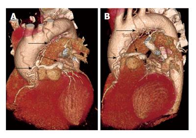
Figure 8 Complex coronary artery fistula.
A and B: 3D volume rendered coronary computed tomography angiography shows an arterial-arterial fistula originating from the proximal right coronary artery and 1st diagonal branch, which terminates in the pulmonary artery. The plexus has an aneurysmal segment that wraps around the anterior aspect of the main pulmonary artery. There is direct communication between the aneurysmal segment and the leftward aspect of the proximal MPA. Also, there are multiple smaller branches around the aortic root, MPA, ascending aorta, and transverse arch. A branch originating from the aortic arch continuous with a small 1.8-cm contrast-filled cavity adjacent to the aortic arch, between the trachea and aortic arch.
- Citation: Ghandour A, Rajiah P. Unusual fistulas and connections in the cardiovascular system: A pictorial review. World J Radiol 2014; 6(5): 169-176
- URL: https://www.wjgnet.com/1949-8470/full/v6/i5/169.htm
- DOI: https://dx.doi.org/10.4329/wjr.v6.i5.169


