Copyright
©2014 Baishideng Publishing Group Inc.
World J Radiol. May 28, 2014; 6(5): 169-176
Published online May 28, 2014. doi: 10.4329/wjr.v6.i5.169
Published online May 28, 2014. doi: 10.4329/wjr.v6.i5.169
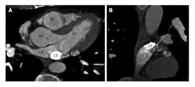
Figure 7 Left circumflex coronary artery to persistent left superior vena cava fistula.
A: Axial coronary computed tomography angiography (CTA) image shows a fistulous connection (F) between left circumflex artery and a persistent left left circumflex coronary artery (LS) which eventually drained into the coronary sinus; B: Sagittal coronary CTA reconstructed images shows fistula (arrow) between the left circumflex artery and persistent left superior vena cava (LS). AO: Aorta; LA: Left atrium.
- Citation: Ghandour A, Rajiah P. Unusual fistulas and connections in the cardiovascular system: A pictorial review. World J Radiol 2014; 6(5): 169-176
- URL: https://www.wjgnet.com/1949-8470/full/v6/i5/169.htm
- DOI: https://dx.doi.org/10.4329/wjr.v6.i5.169


