Copyright
©2014 Baishideng Publishing Group Inc.
World J Radiol. May 28, 2014; 6(5): 169-176
Published online May 28, 2014. doi: 10.4329/wjr.v6.i5.169
Published online May 28, 2014. doi: 10.4329/wjr.v6.i5.169
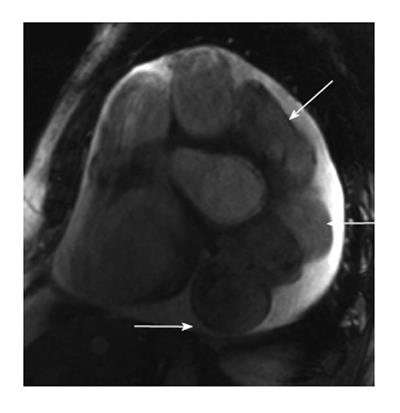
Figure 5 Magnetic resonance imaging appearances of coronary artery fistula.
Short axis steady state free precession magnetic resonance imaging image shows dilated, tortuous left anterior descending artery draining into the coronary sinus. There is also a small circumferential pericardial effusion.
- Citation: Ghandour A, Rajiah P. Unusual fistulas and connections in the cardiovascular system: A pictorial review. World J Radiol 2014; 6(5): 169-176
- URL: https://www.wjgnet.com/1949-8470/full/v6/i5/169.htm
- DOI: https://dx.doi.org/10.4329/wjr.v6.i5.169


