Copyright
©2014 Baishideng Publishing Group Inc.
World J Radiol. May 28, 2014; 6(5): 169-176
Published online May 28, 2014. doi: 10.4329/wjr.v6.i5.169
Published online May 28, 2014. doi: 10.4329/wjr.v6.i5.169
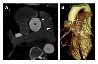
Figure 1 Coronary to pulmonary artery fistula.
A: Axial contrast enhanced coronary computed tomography angiography shows left anterior descending (LAD) artery (long arrow) and right coronary artery (arrowhead) fistulas draining into the pulmonary artery. A jet of contrast (short arrow) is seen emptying into the pulmonary artery; B: Three-dimensional volume rendered computed tomography image shows the LAD coronary artery crossing anterior to the pulmonary artery (PA) to drain into it. AO: Aorta.
- Citation: Ghandour A, Rajiah P. Unusual fistulas and connections in the cardiovascular system: A pictorial review. World J Radiol 2014; 6(5): 169-176
- URL: https://www.wjgnet.com/1949-8470/full/v6/i5/169.htm
- DOI: https://dx.doi.org/10.4329/wjr.v6.i5.169


