Copyright
©2014 Baishideng Publishing Group Inc.
World J Radiol. May 28, 2014; 6(5): 148-159
Published online May 28, 2014. doi: 10.4329/wjr.v6.i5.148
Published online May 28, 2014. doi: 10.4329/wjr.v6.i5.148
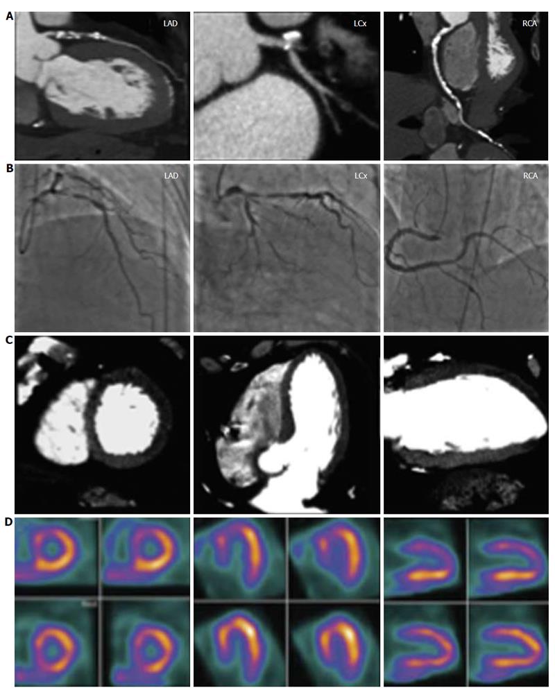
Figure 6 A complete CORE320 imaging data set for a 64-year-old male without prior history of coronary artery disease with chest pain symptoms[50].
The left anterior descending coronary artery revealed a 96% diameter stenosis by computed tomography angiography (CTA) (row A) and an 85% diameter stenosis by invasive coronary angiography (ICA) (row B). The computed tomography myocardial perfusion (CTP) (row C) study revealed a mild defect in the distal anteroseptal wall, and moderate defects in the basal anteroseptal, the basal anterior, the distal anterior, and apical walls, while the single photon emission computed tomography (SPECT) (row D) study revealed moderate defects in the distal anterior, the distal anteroseptal, the basal anteroseptal and apical walls. The left circumflex artery revealed an 87% diameter stenosis by CTA, a 79% diameter stenosis by ICA, mild defects in the distal inferoseptal and distal inferolateral walls, and moderate defects in the distal anterolateral and distal anterior walls by CTP, and a moderate defect in the distal anterior wall by SPECT. The right coronary artery revealed a 60% diameter stenosis by CTA, a 77% diameter stenosis by ICA, a mild defect in the distal inferoseptal wall by CTP, and no myocardial perfusion defects by SPECT.
- Citation: Sato A. Coronary plaque imaging by coronary computed tomography angiography. World J Radiol 2014; 6(5): 148-159
- URL: https://www.wjgnet.com/1949-8470/full/v6/i5/148.htm
- DOI: https://dx.doi.org/10.4329/wjr.v6.i5.148


