Copyright
©2014 Baishideng Publishing Group Co.
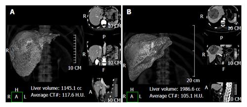
Figure 1 Using a semi-automatic method liver analysis application provides a 3D and a multi-planar reconstruction of the liver.
A case of hilar cholangiocarcinoma involving the left hepatic duct, with marked hypotrophy of the left lobe (type IIIb according to the Bismuth-Corlette classification) (A) and a case of hepatocarcinoma in segments 4-5-8 (B) are shown.
- Citation: D’Onofrio M, De Robertis R, Demozzi E, Crosara S, Canestrini S, Pozzi Mucelli R. Liver volumetry: Is imaging reliable? Personal experience and review of the literature. World J Radiol 2014; 6(4): 62-71
- URL: https://www.wjgnet.com/1949-8470/full/v6/i4/62.htm
- DOI: https://dx.doi.org/10.4329/wjr.v6.i4.62


