Copyright
©2014 Baishideng Publishing Group Co.
World J Radiol. Apr 28, 2014; 6(4): 106-115
Published online Apr 28, 2014. doi: 10.4329/wjr.v6.i4.106
Published online Apr 28, 2014. doi: 10.4329/wjr.v6.i4.106
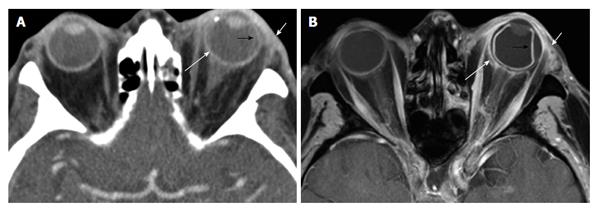
Figure 9 Periscleritic orbital inflammatory disease.
Eighty-seven-year-old immunocompromised man with left eye pain and ordering indication of “cellulitis”. A: Axial contrast-enhanced CT shows mild infiltration of the left periorbital fat (short white arrow). There is also periscleral edema (long white arrow), and subtle high density along the temporal surface of the globe that is suggestive of a subchoroidal fluid collection (black arrow); B: Axial fat-suppressed contrast-enhanced T1 shows these findings more conspicuously. Note that the elevated choroid layer (black arrow) extends anteriorly to the region of the ciliary body. Periscleral edema (long white arrow) extending to Tenon’s capsule is better seen. CT: Computed tomography.
- Citation: Pakdaman MN, Sepahdari AR, Elkhamary SM. Orbital inflammatory disease: Pictorial review and differential diagnosis. World J Radiol 2014; 6(4): 106-115
- URL: https://www.wjgnet.com/1949-8470/full/v6/i4/106.htm
- DOI: https://dx.doi.org/10.4329/wjr.v6.i4.106


