Copyright
©2014 Baishideng Publishing Group Co.
World J Radiol. Apr 28, 2014; 6(4): 106-115
Published online Apr 28, 2014. doi: 10.4329/wjr.v6.i4.106
Published online Apr 28, 2014. doi: 10.4329/wjr.v6.i4.106
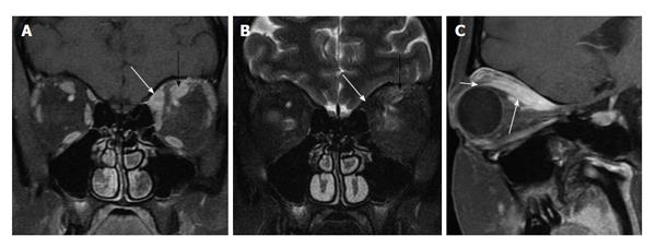
Figure 4 Myositic pseudotumor.
A: Coronal fat-suppressed contrast-enhanced T1 shows enlarged left superior oblique (white arrow) and superior rectus (black arrow) muscles, and mild infiltration of the surrounding fat; B: Coronal fat-suppressed T2 shows low signal in the superior oblique muscle (white arrow), suggesting a more chronic, burned out process, whereas the superior rectus muscle (black arrow) shows brighter signal, indicative of a more acute process; C: Parasagittal oblique fat-suppressed contrast-enhanced T1 shows an enlarged superior rectus muscle belly (long arrow). The tendinous insertion (short arrow) is uncharacteristically spared by this process. Nevertheless, unilateral disease, infiltration of the surrounding fat, and early involvement of the superior oblique muscle indicate pseudotumor ahead of thyroid eye disease.
- Citation: Pakdaman MN, Sepahdari AR, Elkhamary SM. Orbital inflammatory disease: Pictorial review and differential diagnosis. World J Radiol 2014; 6(4): 106-115
- URL: https://www.wjgnet.com/1949-8470/full/v6/i4/106.htm
- DOI: https://dx.doi.org/10.4329/wjr.v6.i4.106


