Copyright
©2014 Baishideng Publishing Group Co.
World J Radiol. Apr 28, 2014; 6(4): 106-115
Published online Apr 28, 2014. doi: 10.4329/wjr.v6.i4.106
Published online Apr 28, 2014. doi: 10.4329/wjr.v6.i4.106
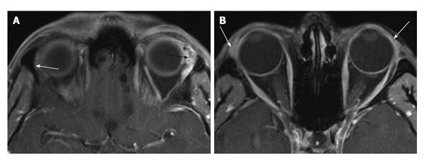
Figure 2 Adenoid cystic carcinoma of the lacrimal gland.
A: Axial T1 non-enhanced MRI showing an enlarged, heterogeneous left lacrimal gland (black arrow). Compare this to the contralateral normal gland (white arrow); B: Axial T1 non-enhanced MRI showing sparing of the palpebral lobe (arrows). MRI: Magnetic resonance imaging.
- Citation: Pakdaman MN, Sepahdari AR, Elkhamary SM. Orbital inflammatory disease: Pictorial review and differential diagnosis. World J Radiol 2014; 6(4): 106-115
- URL: https://www.wjgnet.com/1949-8470/full/v6/i4/106.htm
- DOI: https://dx.doi.org/10.4329/wjr.v6.i4.106


