Copyright
©2014 Baishideng Publishing Group Co.
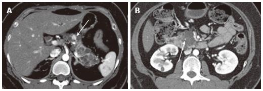
Figure 14 A 43-year-old female with von Hippel-Lindau disease.
A: The arterial phase of an axial contrast enhanced-computed tomography demonstrates numerous cystic lesions in the body and tail of the pancreas. There is a solid hypervascular lesion in the pancreatic body (arrow), representing neuroendocrine tumor; B: A multilocular cystic lesion in the pancreatic head consists of a punctate central calcification (arrow) with microcystic (right side) and macrocystic (left side) components, representing serous cystic neoplasm.
- Citation: Ishigami K, Nishie A, Asayama Y, Ushijima Y, Takayama Y, Fujita N, Takahata S, Ohtsuka T, Ito T, Igarashi H, Ikari S, Metz CM, Honda H. Imaging pitfalls of pancreatic serous cystic neoplasm and its potential mimickers. World J Radiol 2014; 6(3): 36-47
- URL: https://www.wjgnet.com/1949-8470/full/v6/i3/36.htm
- DOI: https://dx.doi.org/10.4329/wjr.v6.i3.36


