Copyright
©2014 Baishideng Publishing Group Co.
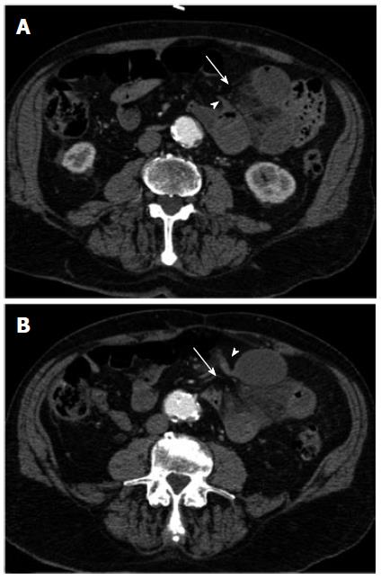
Figure 4 Multi-detector contrast-enhanced computed tomography.
At retrospective analysis, a beak-like appearance (arrow-heads) of the closely apposed afferent (A) and efferent (B) jejunal loops can be appreciated along with the hernial orifice (arrow) in the periphery of the omentum. The peripheral location of the herniated loops and the absence of an overlying omental fat can also be appreciated.
- Citation: Camera L, Gennaro AD, Longobardi M, Masone S, Calabrese E, Vecchio WD, Persico G, Salvatore M. A spontaneous strangulated transomental hernia: Prospective and retrospective multi-detector computed tomography findings. World J Radiol 2014; 6(2): 26-30
- URL: https://www.wjgnet.com/1949-8470/full/v6/i2/26.htm
- DOI: https://dx.doi.org/10.4329/wjr.v6.i2.26


