Copyright
©2014 Baishideng Publishing Group Co.
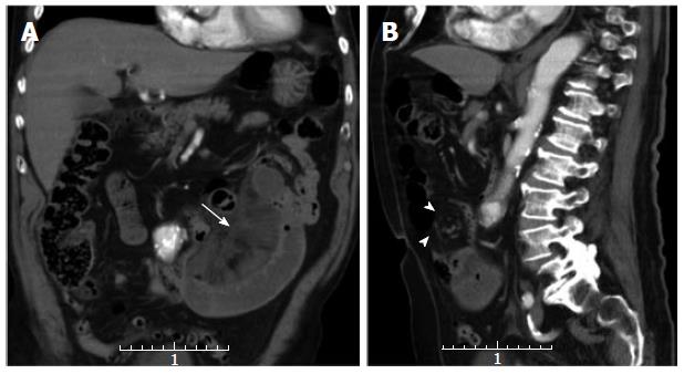
Figure 3 Multi-detector contrast-enhanced computed tomography.
Coronal (A) and sagittal (B) reformatted 5 mm thick images are shown. In (A) a cluster of ischemic jejunal loops is depicted in the left flank along with mesenteric fluid (arrow). In (B) swirling of the mesenteric vessels (arrow-heads) is depicted. The computed tomography finding (whirl sign) was prospectively considered consistent with a small bowel volvulus which was not confirmed at surgery.
- Citation: Camera L, Gennaro AD, Longobardi M, Masone S, Calabrese E, Vecchio WD, Persico G, Salvatore M. A spontaneous strangulated transomental hernia: Prospective and retrospective multi-detector computed tomography findings. World J Radiol 2014; 6(2): 26-30
- URL: https://www.wjgnet.com/1949-8470/full/v6/i2/26.htm
- DOI: https://dx.doi.org/10.4329/wjr.v6.i2.26


