Copyright
©2014 Baishideng Publishing Group Inc.
World J Radiol. Dec 28, 2014; 6(12): 924-927
Published online Dec 28, 2014. doi: 10.4329/wjr.v6.i12.924
Published online Dec 28, 2014. doi: 10.4329/wjr.v6.i12.924
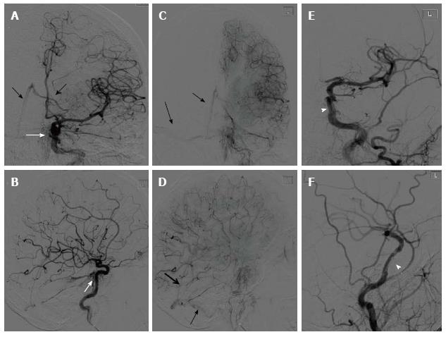
Figure 1 Left internal carotid artery angiogram in the AP and lateral views (arterial phase: A and B; capillary phase: C and D; and following the deployment of the Jostent Graftmaster: E and F).
White arrows show the contrast extravasations at the cavernous segment pointing posteriorly with early venous filling to the internal cerebral vein, straight and lateral venous sinuses (black arrows). White arrow heads shows Jostent Graftmaster in position with decreased contrast extravasations.
- Citation: Allam H, Callison RC, Scodary D, Alawi A, Hogan DW, Alshekhlee A. Traumatic carotid-rosenthal fistula treated with Jostent Graftmaster. World J Radiol 2014; 6(12): 924-927
- URL: https://www.wjgnet.com/1949-8470/full/v6/i12/924.htm
- DOI: https://dx.doi.org/10.4329/wjr.v6.i12.924


