Copyright
©2014 Baishideng Publishing Group Inc.
World J Radiol. Dec 28, 2014; 6(12): 919-923
Published online Dec 28, 2014. doi: 10.4329/wjr.v6.i12.919
Published online Dec 28, 2014. doi: 10.4329/wjr.v6.i12.919
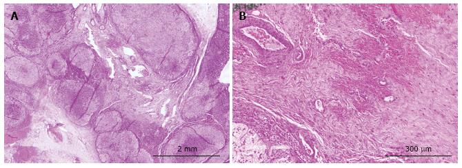
Figure 4 Low power view (A), showing nodular adrenal gland with intervening fibrotic areas.
High power picture (B) demonstrating a hypocellular spindle cell proliferation. Areas of hemorrhage as well as the presence of thick walled blood vessels were also noted.
- Citation: Wassal EY, Habra MA, Vicens R, Rao P, Elsayes KM. Ovarian thecal metaplasia of the adrenal gland in association with Beckwith-Wiedemann syndrome. World J Radiol 2014; 6(12): 919-923
- URL: https://www.wjgnet.com/1949-8470/full/v6/i12/919.htm
- DOI: https://dx.doi.org/10.4329/wjr.v6.i12.919


