Copyright
©2014 Baishideng Publishing Group Inc.
World J Radiol. Dec 28, 2014; 6(12): 913-918
Published online Dec 28, 2014. doi: 10.4329/wjr.v6.i12.913
Published online Dec 28, 2014. doi: 10.4329/wjr.v6.i12.913
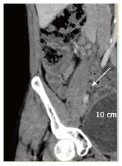
Figure 5 Thirty-five-year-old woman with history of asymptomatic hematuria.
Normal appendix was not visualized on non-contrast study due to fluid distended adjacent bowel and paucity of intra-abdominal fat. Above shown post intravenous contrast computed tomography of the same patient later helped identify a fluid-filled appendix of 6.0 mm diameter (arrow).
- Citation: Yaqoob J, Idris M, Alam MS, Kashif N. Can outer-to-outer diameter be used alone in diagnosing appendicitis on 128-slice MDCT? World J Radiol 2014; 6(12): 913-918
- URL: https://www.wjgnet.com/1949-8470/full/v6/i12/913.htm
- DOI: https://dx.doi.org/10.4329/wjr.v6.i12.913


