Copyright
©2014 Baishideng Publishing Group Inc.
World J Radiol. Dec 28, 2014; 6(12): 890-894
Published online Dec 28, 2014. doi: 10.4329/wjr.v6.i12.890
Published online Dec 28, 2014. doi: 10.4329/wjr.v6.i12.890
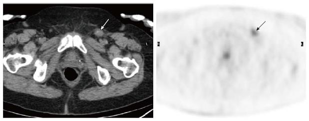
Figure 2 Axial image of F18-fluoro-2-deoxy-D-glucose positron emission tomography/computed tomography obtained 5 mo postoperatively in a 68-year-old woman with history of vulvar squamous cell carcinoma.
Compared to preoperative image, there was a new 1.5 cm left inguinal lymph node with increased uptake (SUV 4.9, arrows). Incisional biopsy of the node suggested lymphadenitis. SUV: Standardized uptake value.
- Citation: Liu Y. Postoperative reactive lymphadenitis: A potential cause of false-positive FDG PET/CT. World J Radiol 2014; 6(12): 890-894
- URL: https://www.wjgnet.com/1949-8470/full/v6/i12/890.htm
- DOI: https://dx.doi.org/10.4329/wjr.v6.i12.890


