Copyright
©2014 Baishideng Publishing Group Inc.
World J Radiol. Nov 28, 2014; 6(11): 886-889
Published online Nov 28, 2014. doi: 10.4329/wjr.v6.i11.886
Published online Nov 28, 2014. doi: 10.4329/wjr.v6.i11.886
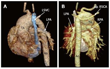
Figure 2 Volume rendered 3D images (A and B) clearly demonstrates correlation between these abnormal vessels and origins.
TA: Truncus arteriosus; RPA: Right pulmonary artery; LPA: Left pulmonary artery; Ao: Descending aorta; LSVC: Left superior vena cava; IVC: Inferior vena cava; Ab RSCA: Right aberrant subclavian artery.
- Citation: Koplay M, Cimen D, Sivri M, Güvenc O, Arslan D, Nayman A, Oran B. Truncus arteriosus: Diagnosis with dual-source computed tomography angiography and low radiation dose. World J Radiol 2014; 6(11): 886-889
- URL: https://www.wjgnet.com/1949-8470/full/v6/i11/886.htm
- DOI: https://dx.doi.org/10.4329/wjr.v6.i11.886


