Copyright
©2014 Baishideng Publishing Group Inc.
World J Radiol. Nov 28, 2014; 6(11): 865-873
Published online Nov 28, 2014. doi: 10.4329/wjr.v6.i11.865
Published online Nov 28, 2014. doi: 10.4329/wjr.v6.i11.865
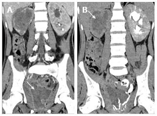
Figure 20 Eosinophilic cystitis.
A 25 years old man who presented with hematuria and worsening irritative symptoms over past one year. Clinical suspicion was that of a bladder malignancy. A: Coronal reformatted CECT in nephrographic phase shows diffuse mass like bladder wall thickening and irregularity with air specks in the wall. Mass like soft tissue is replacing entire right kidney with perinephric spread; B: Delayed coronal CECT shows opacification of rectum through a fistulous communication (arrow). Note made of striated nephrogram in left kidney suggesting ongoing acute inflammatory process. Biopsy revealed eosinophilic infiltration and fibrosis within the bladder wall with no evidence of malignancy. CECT: Contrast-enhanced computed tomography.
- Citation: Das CJ, Ahmad Z, Sharma S, Gupta AK. Multimodality imaging of renal inflammatory lesions. World J Radiol 2014; 6(11): 865-873
- URL: https://www.wjgnet.com/1949-8470/full/v6/i11/865.htm
- DOI: https://dx.doi.org/10.4329/wjr.v6.i11.865


