Copyright
©2014 Baishideng Publishing Group Inc.
World J Radiol. Nov 28, 2014; 6(11): 865-873
Published online Nov 28, 2014. doi: 10.4329/wjr.v6.i11.865
Published online Nov 28, 2014. doi: 10.4329/wjr.v6.i11.865
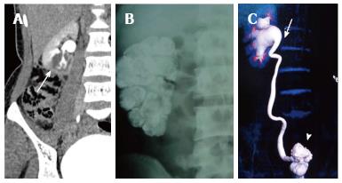
Figure 17 Renal tuberculosis.
A: Delayed CECT shows a cavitation at the lower pole of right kidney communicating with the PCS. This finding is fairly typical of GU TB. This adolescent male was a known case of pulmonary tuberculosis; B: Plain abdominal radiograph in a different patient shows diffuse parenchymal calcification of right kidney suggestive endstage autonephrectomy or putty kidney; C: Volume rendered technique image of delayed phase CECT shows a contracted thimble bladder (arrowhead), hiked up right pelvis (arrow) and hydroureteronephrosis. This patient had acid fast bacilli cultured from urine. PCS: Pelvicalyceal system; CECT: Contrast-enhanced computed tomography; TB: Tuberculosis.
- Citation: Das CJ, Ahmad Z, Sharma S, Gupta AK. Multimodality imaging of renal inflammatory lesions. World J Radiol 2014; 6(11): 865-873
- URL: https://www.wjgnet.com/1949-8470/full/v6/i11/865.htm
- DOI: https://dx.doi.org/10.4329/wjr.v6.i11.865


