Copyright
©2014 Baishideng Publishing Group Inc.
World J Radiol. Nov 28, 2014; 6(11): 865-873
Published online Nov 28, 2014. doi: 10.4329/wjr.v6.i11.865
Published online Nov 28, 2014. doi: 10.4329/wjr.v6.i11.865
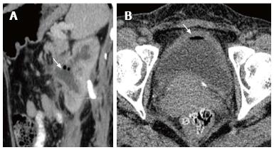
Figure 15 Emphysematous pyelitis and cystitis.
A: Para-sagittal reformatted CECT of a 42 years old diabetic lady showing air within dilated PCS with surrounding inflammatory changes; B: Axial CECT shows bladder wall thickening and air within the bladder lumen. PCS: Pelvicalyceal system; CECT: Contrast-enhanced computed tomography.
- Citation: Das CJ, Ahmad Z, Sharma S, Gupta AK. Multimodality imaging of renal inflammatory lesions. World J Radiol 2014; 6(11): 865-873
- URL: https://www.wjgnet.com/1949-8470/full/v6/i11/865.htm
- DOI: https://dx.doi.org/10.4329/wjr.v6.i11.865


