Copyright
©2014 Baishideng Publishing Group Inc.
World J Radiol. Oct 28, 2014; 6(10): 840-845
Published online Oct 28, 2014. doi: 10.4329/wjr.v6.i10.840
Published online Oct 28, 2014. doi: 10.4329/wjr.v6.i10.840
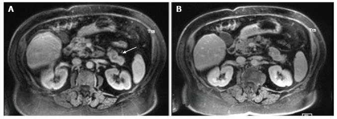
Figure 3 Contrast-enhanced magnetic resonance.
Gadolinium-enhanced (0.1 mmol/kg) VIBE T1 Fat-suppressed 3 mm thick images are shown in both the arterial (A) and delayed (B) phase. Despite motion artifacts arterial phase image (A) shows an inhomogeneously enhancing lesion (3.7 cm × 1.7 cm) at the level of the pancreatic tail (arrow). The lesion exhibits a rim of peripheral enhancement (arrow-head) in the delayed phase (B).
- Citation: Camera L, Severino R, Faggiano A, Masone S, Mansueto G, Maurea S, Fonti R, Salvatore M. Contrast enhanced multi-detector CT and MR findings of a well-differentiated pancreatic vipoma. World J Radiol 2014; 6(10): 840-845
- URL: https://www.wjgnet.com/1949-8470/full/v6/i10/840.htm
- DOI: https://dx.doi.org/10.4329/wjr.v6.i10.840


