Copyright
©2014 Baishideng Publishing Group Inc.
World J Radiol. Oct 28, 2014; 6(10): 808-825
Published online Oct 28, 2014. doi: 10.4329/wjr.v6.i10.808
Published online Oct 28, 2014. doi: 10.4329/wjr.v6.i10.808
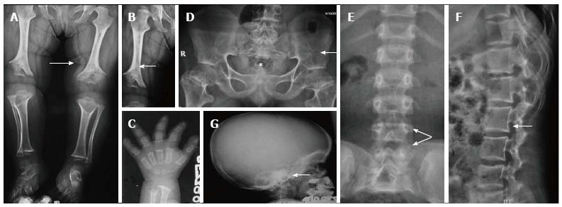
Figure 8 Achondroplasia.
Radiograph of lower limbs (A, B) shows bilateral rhizomelic shortening with metaphyseal flaring (arrow, A) and chevron deformity in femur (arrow, B). Note trident hand appearance in (C). Radiograph of pelvis (D) shows short and broad pelvis (*), horizontal acetabuli (arrow) and round iliac wings. Radiographs of spine (E, F) show narrow interpedicular distance in lumbar spine (arrow, E) and posterior scalloping and thick, short pedicles (arrow, F). Radiograph of skull (G) shows enlarged cranial vault with narrowed foramen magnum (arrow).
- Citation: Panda A, Gamanagatti S, Jana M, Gupta AK. Skeletal dysplasias: A radiographic approach and review of common non-lethal skeletal dysplasias. World J Radiol 2014; 6(10): 808-825
- URL: https://www.wjgnet.com/1949-8470/full/v6/i10/808.htm
- DOI: https://dx.doi.org/10.4329/wjr.v6.i10.808


