Copyright
©2014 Baishideng Publishing Group Inc.
World J Radiol. Oct 28, 2014; 6(10): 808-825
Published online Oct 28, 2014. doi: 10.4329/wjr.v6.i10.808
Published online Oct 28, 2014. doi: 10.4329/wjr.v6.i10.808
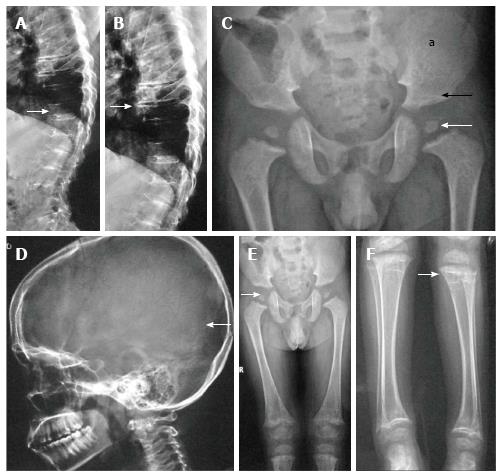
Figure 1 Spondyloepiphyseal dysplasia congenita.
Lateral radiographs of dorsolumbar spine show platyspondyly (arrow, A) with severely reduced intervertebral disc spaces (arrow, B). Radiograph of pelvis (C) shows small femoral epiphyses (white arrow), horizontal acetabuli (black arrow) and short iliac wings (a). Radiograph of skull (D) shows relatively enlarged calvarium (arrow). Radiographs of lower limbs (E, F) show relatively short femurs and small epiphyses with secondary metaphyseal irregularity (arrow, F).
- Citation: Panda A, Gamanagatti S, Jana M, Gupta AK. Skeletal dysplasias: A radiographic approach and review of common non-lethal skeletal dysplasias. World J Radiol 2014; 6(10): 808-825
- URL: https://www.wjgnet.com/1949-8470/full/v6/i10/808.htm
- DOI: https://dx.doi.org/10.4329/wjr.v6.i10.808


