Copyright
©2014 Baishideng Publishing Group Inc.
World J Radiol. Oct 28, 2014; 6(10): 794-807
Published online Oct 28, 2014. doi: 10.4329/wjr.v6.i10.794
Published online Oct 28, 2014. doi: 10.4329/wjr.v6.i10.794
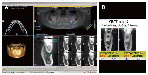
Figure 3 Cone beam computed tomography.
A: A cone beam computed tomography scan gives a three-dimensional view of the area of interest. In this case, the periapical lesion is being evaluated; B: The image gives values in Hounsfield unit of cementum and alveolar bone density to measure post-treatment healing. CBCT: Cone beam computed tomography.
- Citation: Shah N, Bansal N, Logani A. Recent advances in imaging technologies in dentistry. World J Radiol 2014; 6(10): 794-807
- URL: https://www.wjgnet.com/1949-8470/full/v6/i10/794.htm
- DOI: https://dx.doi.org/10.4329/wjr.v6.i10.794


