Copyright
©2014 Baishideng Publishing Group Inc.
World J Radiol. Oct 28, 2014; 6(10): 779-793
Published online Oct 28, 2014. doi: 10.4329/wjr.v6.i10.779
Published online Oct 28, 2014. doi: 10.4329/wjr.v6.i10.779
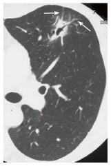
Figure 27 Pulmonary malignant lymphoma (mucosa associated lymphoid tissue lymphoma) in a woman in her 30s.
Thin-section computed tomography shows a focal consolidation with dilated air bronchograms in the lingula of the left upper lobe. Note mild thickening of the vessels penetrating the consolidation (arrows), suggestive of infiltrative growth of malignant lymphoma along the vessels.
- Citation: Nambu A, Ozawa K, Kobayashi N, Tago M. Imaging of community-acquired pneumonia: Roles of imaging examinations, imaging diagnosis of specific pathogens and discrimination from noninfectious diseases. World J Radiol 2014; 6(10): 779-793
- URL: https://www.wjgnet.com/1949-8470/full/v6/i10/779.htm
- DOI: https://dx.doi.org/10.4329/wjr.v6.i10.779


