Copyright
©2014 Baishideng Publishing Group Inc.
World J Radiol. Oct 28, 2014; 6(10): 779-793
Published online Oct 28, 2014. doi: 10.4329/wjr.v6.i10.779
Published online Oct 28, 2014. doi: 10.4329/wjr.v6.i10.779
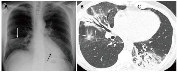
Figure 23 Cryptogenic organizing pneumonia in a woman in her 50s.
A: Chest radiograph shows a consolidation in the right lower lung field with depression of the right minor fissure suggestive of volume loss of the middle lobe (white arrow). Retrocardiac consolidation is marginally seen (black arrow); B, C: Thin-section computed tomography of the right lung (B) and left lung (C) demonstrate consolidations with air bronchograms in both lungs. Note that the bronchi within the consolidations are mildly dilated and that the consolidations have concaved margins (arrows), suggesting organization of the disease.
- Citation: Nambu A, Ozawa K, Kobayashi N, Tago M. Imaging of community-acquired pneumonia: Roles of imaging examinations, imaging diagnosis of specific pathogens and discrimination from noninfectious diseases. World J Radiol 2014; 6(10): 779-793
- URL: https://www.wjgnet.com/1949-8470/full/v6/i10/779.htm
- DOI: https://dx.doi.org/10.4329/wjr.v6.i10.779


