Copyright
©2014 Baishideng Publishing Group Inc.
World J Radiol. Oct 28, 2014; 6(10): 779-793
Published online Oct 28, 2014. doi: 10.4329/wjr.v6.i10.779
Published online Oct 28, 2014. doi: 10.4329/wjr.v6.i10.779
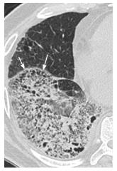
Figure 22 Pneumonia on a background of pulmonary emphysema mimicking honeycombing in a man in his 60s (causative pathogen unknown).
Thin-section computed tomography shows an extensive area of ground-glass opacity intermingled with consolidation in the right lower lobe. Pseudohoneycombing is seen along the interlobar fissure (arrows).
- Citation: Nambu A, Ozawa K, Kobayashi N, Tago M. Imaging of community-acquired pneumonia: Roles of imaging examinations, imaging diagnosis of specific pathogens and discrimination from noninfectious diseases. World J Radiol 2014; 6(10): 779-793
- URL: https://www.wjgnet.com/1949-8470/full/v6/i10/779.htm
- DOI: https://dx.doi.org/10.4329/wjr.v6.i10.779


