Copyright
©2014 Baishideng Publishing Group Inc.
World J Radiol. Oct 28, 2014; 6(10): 779-793
Published online Oct 28, 2014. doi: 10.4329/wjr.v6.i10.779
Published online Oct 28, 2014. doi: 10.4329/wjr.v6.i10.779
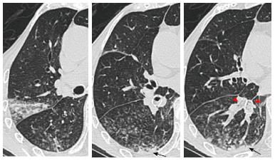
Figure 19 Aspiration pneumonia in a woman in her 70s (causative pathogen unknown).
Thin-section computed tomography images show centrilobular nodules with surrounding ground-glass opacities and subpleural non-segmental consolidations (arrows) at the dorsal portions of the right lung. Note that the lumens of segmental bronchi are filled with aspirated materials (arrow heads).
- Citation: Nambu A, Ozawa K, Kobayashi N, Tago M. Imaging of community-acquired pneumonia: Roles of imaging examinations, imaging diagnosis of specific pathogens and discrimination from noninfectious diseases. World J Radiol 2014; 6(10): 779-793
- URL: https://www.wjgnet.com/1949-8470/full/v6/i10/779.htm
- DOI: https://dx.doi.org/10.4329/wjr.v6.i10.779


