Copyright
©2014 Baishideng Publishing Group Inc.
World J Radiol. Oct 28, 2014; 6(10): 779-793
Published online Oct 28, 2014. doi: 10.4329/wjr.v6.i10.779
Published online Oct 28, 2014. doi: 10.4329/wjr.v6.i10.779
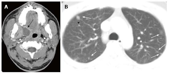
Figure 17 Pulmonary septic emboli from paratonsillar abscess in a man in his 20s.
A: Enhanced computed tomography (CT) at the level of oropharynx shows an abscess at the right paratonsillar region (arrow); B: Chest CT with a 5 mm slice thickness reveals small nodules in both lungs (arrows), some of which are in contact with the periphery of pulmonary vessels (arrow head).
- Citation: Nambu A, Ozawa K, Kobayashi N, Tago M. Imaging of community-acquired pneumonia: Roles of imaging examinations, imaging diagnosis of specific pathogens and discrimination from noninfectious diseases. World J Radiol 2014; 6(10): 779-793
- URL: https://www.wjgnet.com/1949-8470/full/v6/i10/779.htm
- DOI: https://dx.doi.org/10.4329/wjr.v6.i10.779


