Copyright
©2014 Baishideng Publishing Group Inc.
World J Radiol. Oct 28, 2014; 6(10): 779-793
Published online Oct 28, 2014. doi: 10.4329/wjr.v6.i10.779
Published online Oct 28, 2014. doi: 10.4329/wjr.v6.i10.779
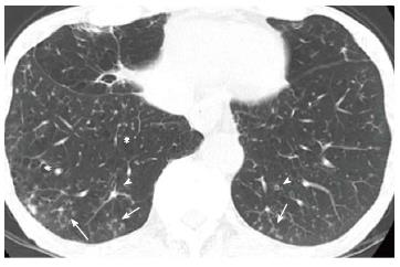
Figure 8 Chronic transbronchial infection (diffuse aspiration bronchiolitis) in a man in his 70s.
This patient had a history of esophageal carcinoma and associated repeated aspiration. Thin-section computed tomography at the level of lung base shows centrilobular nodules (arrows) with bronchiectasis (arrow heads). Low attenuation areas suggestive of pulmonary emphysema are also present (*).
- Citation: Nambu A, Ozawa K, Kobayashi N, Tago M. Imaging of community-acquired pneumonia: Roles of imaging examinations, imaging diagnosis of specific pathogens and discrimination from noninfectious diseases. World J Radiol 2014; 6(10): 779-793
- URL: https://www.wjgnet.com/1949-8470/full/v6/i10/779.htm
- DOI: https://dx.doi.org/10.4329/wjr.v6.i10.779


