Copyright
©2014 Baishideng Publishing Group Inc.
World J Radiol. Oct 28, 2014; 6(10): 741-755
Published online Oct 28, 2014. doi: 10.4329/wjr.v6.i10.741
Published online Oct 28, 2014. doi: 10.4329/wjr.v6.i10.741
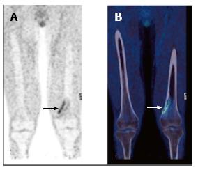
Figure 10 Non–ossifying fibroma (arrow) in the left distal femur of a 17-year-old male patient with Langerhans cell histiocytosis.
FDG PET (A) showing avid uptake, CT (B) showing typical benign radiological features. PET: Positron emission tomography; CT: Computed tomography; FDG: 2-deoxy-2-(18F)fluoro-D-glucose.
- Citation: Freebody J, Wegner EA, Rossleigh MA. 2-deoxy-2-(18F)fluoro-D-glucose positron emission tomography/computed tomography imaging in paediatric oncology. World J Radiol 2014; 6(10): 741-755
- URL: https://www.wjgnet.com/1949-8470/full/v6/i10/741.htm
- DOI: https://dx.doi.org/10.4329/wjr.v6.i10.741


