Copyright
©2014 Baishideng Publishing Group Inc.
World J Radiol. Oct 28, 2014; 6(10): 741-755
Published online Oct 28, 2014. doi: 10.4329/wjr.v6.i10.741
Published online Oct 28, 2014. doi: 10.4329/wjr.v6.i10.741
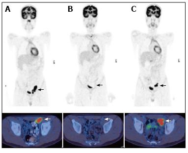
Figure 1 A 13 year-old male with nodular sclerosing Hodgkin’s disease.
PET/CT at staging (A) demonstrated disease in the left external iliac region. After completion of chemotherapy 4 mo later (B) there was a complete metabolic response with no activity in the residual lymph node mass. PET/CT performed 6 mo later (C) for surveillance demonstrated a recurrence at the same site. PET/CT: Positron emission tomography/computed tomography.
- Citation: Freebody J, Wegner EA, Rossleigh MA. 2-deoxy-2-(18F)fluoro-D-glucose positron emission tomography/computed tomography imaging in paediatric oncology. World J Radiol 2014; 6(10): 741-755
- URL: https://www.wjgnet.com/1949-8470/full/v6/i10/741.htm
- DOI: https://dx.doi.org/10.4329/wjr.v6.i10.741


