Copyright
©2014 Baishideng Publishing Group Co.
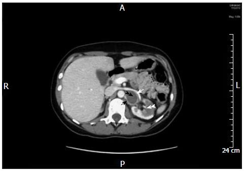
Figure 1 Axial computed tomography scan of the abdomen in the portovenous phase at the level of the kidney.
Black double-arrow head points to hypodense tumor with ring enhancement at the renal hilum. White arrow shows the displaced renal vein; black arrow shows the compressed renal artery. White double-arrow head shows anterior renal infarct.
- Citation: Yehia ZAA, Sayyid RK, Haydar AA. Renal hilar paraganglioma: A case report. World J Radiol 2014; 6(1): 15-17
- URL: https://www.wjgnet.com/1949-8470/full/v6/i1/15.htm
- DOI: https://dx.doi.org/10.4329/wjr.v6.i1.15


