Copyright
©2013 Baishideng Publishing Group Co.
World J Radiol. Aug 28, 2013; 5(8): 328-333
Published online Aug 28, 2013. doi: 10.4329/wjr.v5.i8.328
Published online Aug 28, 2013. doi: 10.4329/wjr.v5.i8.328
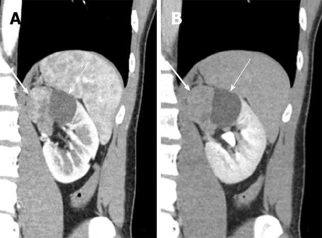
Figure 2 Coronal images of dynamic computer tomography scans show a well-circumscribed, bilobed renal mass in the left renal upper pole with a relatively preserved reniform shape and no definite perirenal fat infiltration or renal sinus invasion or herniation.
The early enhancing solid right half (thick arrow, A) and the non-enhancing cystic left half (thin arrow, A) are apparent. The delayed washout pattern of the enhancing solid portion is evident in the nephrographic phase (thick arrow, B).
- Citation: Yoon JH. Primary renal carcinoid tumor: A rare cystic renal neoplasm. World J Radiol 2013; 5(8): 328-333
- URL: https://www.wjgnet.com/1949-8470/full/v5/i8/328.htm
- DOI: https://dx.doi.org/10.4329/wjr.v5.i8.328


