Copyright
©2013 Baishideng Publishing Group Co.
World J Radiol. Aug 28, 2013; 5(8): 321-324
Published online Aug 28, 2013. doi: 10.4329/wjr.v5.i8.321
Published online Aug 28, 2013. doi: 10.4329/wjr.v5.i8.321
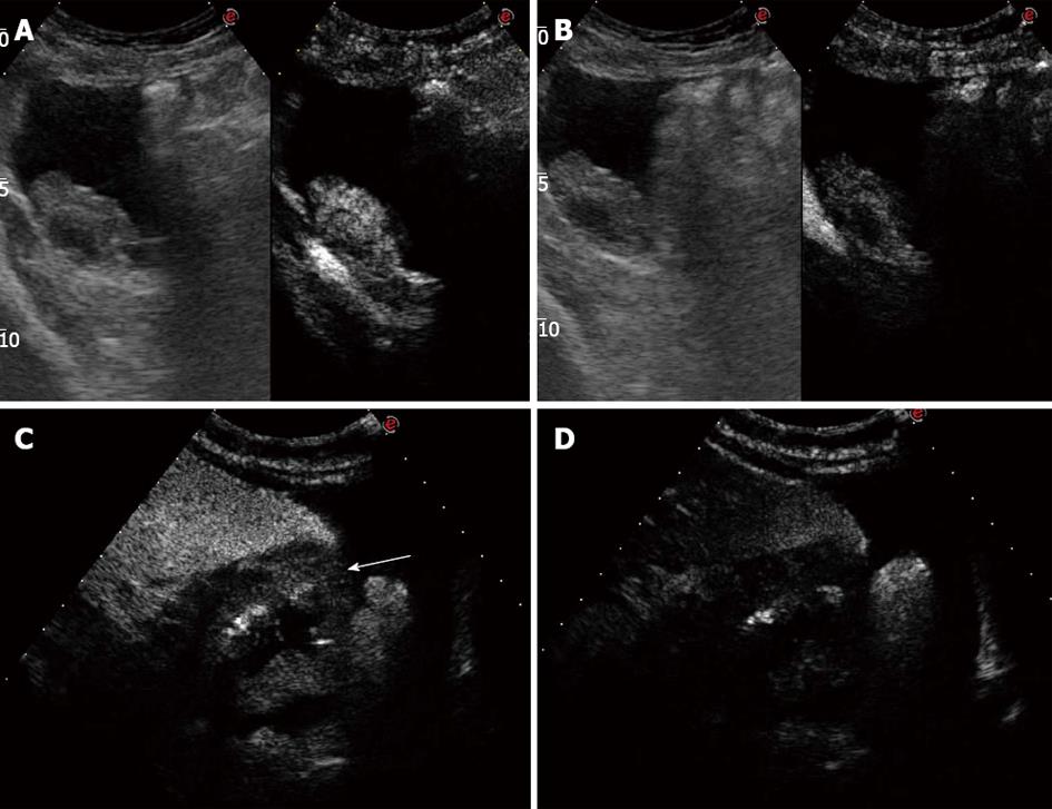
Figure 2 Contrast-enhanced ultrasonographyscans focused.
A: Inhomogeneous contrast enhancement in the arterial phase on the adnexal mass; B: Wash-out in the late venous phase on the adnexal mass; C:Contrast enhancement in the arterial phase on the stomach (arrow); D: marked wash-out in the late venous phase on the stomach.
- Citation: Tombesi P, Vece FD, Ermili F, Fabbian F, Sartori S. Role of ultrasonography and contrast-enhanced ultrasonography in a case of Krukenberg tumor. World J Radiol 2013; 5(8): 321-324
- URL: https://www.wjgnet.com/1949-8470/full/v5/i8/321.htm
- DOI: https://dx.doi.org/10.4329/wjr.v5.i8.321


