Copyright
©2013 Baishideng Publishing Group Co.
World J Radiol. Aug 28, 2013; 5(8): 304-312
Published online Aug 28, 2013. doi: 10.4329/wjr.v5.i8.304
Published online Aug 28, 2013. doi: 10.4329/wjr.v5.i8.304
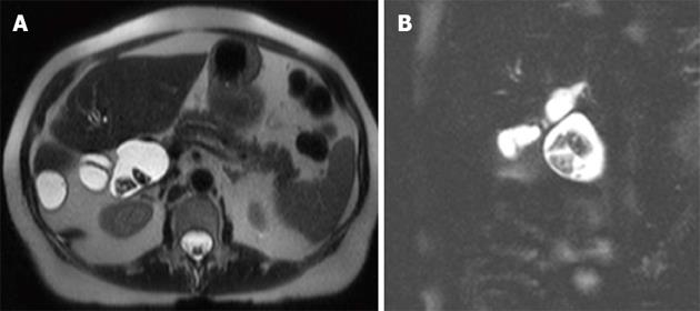
Figure 3 Seventy four-year-old female.
Axial and coronal T2 weighted half-fourier acquisition single-shot turbo spin-echo images showing type IV choledochal cyst with multiple stones in the lumen.
- Citation: Sacher VY, Davis JS, Sleeman D, Casillas J. Role of magnetic resonance cholangiopancreatography in diagnosing choledochal cysts: Case series and review. World J Radiol 2013; 5(8): 304-312
- URL: https://www.wjgnet.com/1949-8470/full/v5/i8/304.htm
- DOI: https://dx.doi.org/10.4329/wjr.v5.i8.304


