Copyright
©2013 Baishideng Publishing Group Co.
World J Radiol. Aug 28, 2013; 5(8): 285-294
Published online Aug 28, 2013. doi: 10.4329/wjr.v5.i8.285
Published online Aug 28, 2013. doi: 10.4329/wjr.v5.i8.285
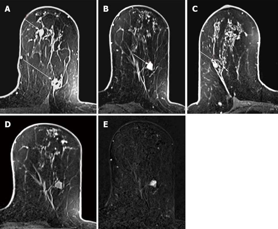
Figure 1 3.
0Tesla axial T1-weighted fat-suppressed contrast enhanced images in 62-year-old woman with newly diagnosed left breast infiltrating ductal carcinoma show the index cancer with signal void due to biopsy marker (long arrow) in the superior lateral quadrant and a second mammographically occult multicentric lesion shown to represent DCIS (short arrow) in the superior medial quadrant (A), an additional multicentric lesion proven to represent an additional infiltrating ductal carcinoma (arrow) in the inferior lateral quadrant (B), and a suspicious contralateral lesion (arrow) confirmed as a benign papilloma (C).
- Citation: Butler RS, Chen C, Vashi R, Hooley RJ, Philpotts LE. 3.0 Tesla vs 1.5 Tesla breast magnetic resonance imaging in newly diagnosed breast cancer patients. World J Radiol 2013; 5(8): 285-294
- URL: https://www.wjgnet.com/1949-8470/full/v5/i8/285.htm
- DOI: https://dx.doi.org/10.4329/wjr.v5.i8.285


