Copyright
©2013 Baishideng Publishing Group Co.
World J Radiol. Aug 28, 2013; 5(8): 275-284
Published online Aug 28, 2013. doi: 10.4329/wjr.v5.i8.275
Published online Aug 28, 2013. doi: 10.4329/wjr.v5.i8.275
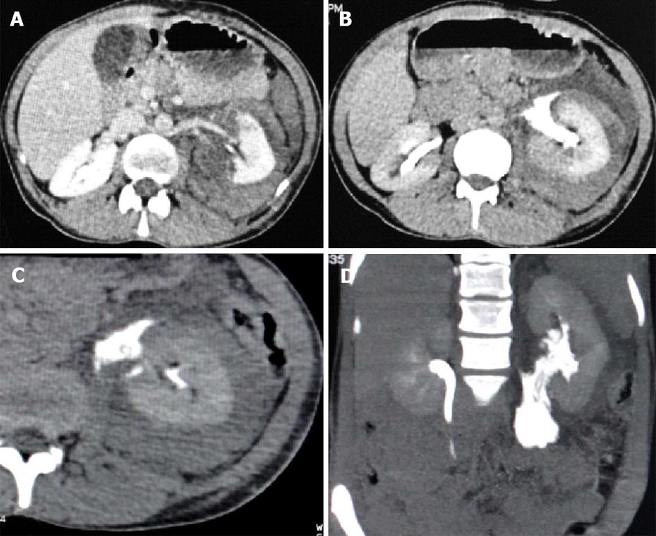
Figure 8 Grade V injury.
Ureteropelvic junction injury (revised stage Grade IV). Contrast enhanced computed tomography images of a 30-year-old female patient who sustained blunt abdominal trauma. A: Axial section showing deep laceration in posteromedial cortex of left kidney involving the hilum with perinephric and perihilar hematoma; B: Excretory phase axial section showing leak of opacified urine from the pelviureteric junction which tracks along the medial aspect of kidney; C: Axial maximum intensity projection (MIP); D: Coronal MIP image showing the extravasated opacified urine from the renal pelvis and tracking in the retroperitoneum along the course of ureter.
- Citation: Dayal M, Gamanagatti S, Kumar A. Imaging in renal trauma. World J Radiol 2013; 5(8): 275-284
- URL: https://www.wjgnet.com/1949-8470/full/v5/i8/275.htm
- DOI: https://dx.doi.org/10.4329/wjr.v5.i8.275


