Copyright
©2013 Baishideng Publishing Group Co.
World J Radiol. Jun 28, 2013; 5(6): 229-240
Published online Jun 28, 2013. doi: 10.4329/wjr.v5.i6.229
Published online Jun 28, 2013. doi: 10.4329/wjr.v5.i6.229
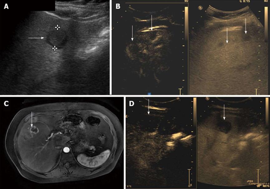
Figure 3 Metastases from neuroendocrine tumor.
A: Hypoechoic target like lesion on B mode ultrasound on routine ultrasound (arrow) B: Contrast enhanced ultrasound (CEUS) shows at least 2 lesions (arrows) with early arterial peripheral hypervascularity with synchronous B mode image of the lesions; C: T1 weighted contrast enhanced axial section of the liver on magnetic resonance imaging with hypervascular peripheral enhancing rim of the lesion in arterial phase (arrow); D: CEUS shows washout on delayed scans (arrow).
- Citation: Laroia ST, Bawa SS, Jain D, Mukund A, Sarin S. Contrast ultrasound in hepatocellular carcinoma at a tertiary liver center: First Indian experience. World J Radiol 2013; 5(6): 229-240
- URL: https://www.wjgnet.com/1949-8470/full/v5/i6/229.htm
- DOI: https://dx.doi.org/10.4329/wjr.v5.i6.229


