Copyright
©2013 Baishideng Publishing Group Co.
World J Radiol. May 28, 2013; 5(5): 220-225
Published online May 28, 2013. doi: 10.4329/wjr.v5.i5.220
Published online May 28, 2013. doi: 10.4329/wjr.v5.i5.220
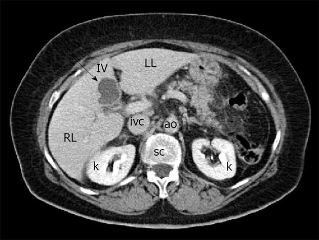
Figure 2 Computed tomography shows the biloma as a hypodense lesion in the IV hepatic segment, characterized by absence of enhancement after administration of intravenous contrast agent (arrow).
RL: Right hepatic lobe; LL: Left hepatic lobe; IV: Fourth hepatic segment; k: Kidney; ivc: Inferior vena cava; ao: Aorta; sc: Spinal column.
- Citation: Tana C, D’Alessandro P, Tartaro A, Tana M, Mezzetti A, Schiavone C. Sonographic assessment of a suspected biloma: A case report and review of the literature. World J Radiol 2013; 5(5): 220-225
- URL: https://www.wjgnet.com/1949-8470/full/v5/i5/220.htm
- DOI: https://dx.doi.org/10.4329/wjr.v5.i5.220


