Copyright
©2013 Baishideng Publishing Group Co.
World J Radiol. May 28, 2013; 5(5): 220-225
Published online May 28, 2013. doi: 10.4329/wjr.v5.i5.220
Published online May 28, 2013. doi: 10.4329/wjr.v5.i5.220
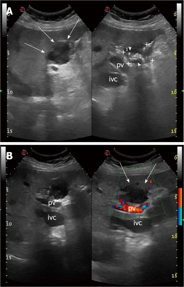
Figure 1 Chronic biloma in a 72-year-old woman: ultrasound findings.
A: Oblique view shows a heterogeneous hypo-anechoic rounded lesion with hyperechoic, calcified walls (arrows), numerous hyperechoic debris generating acustic shadow (arrow heads) and maximum size of 3.89 cm (caliper 1) × 3.42 cm (caliper 2); B: Color Doppler sonogram shows absence of vascularity inside the lesion (arrows). pv: Portal vein; ivc: Inferior vena cava.
- Citation: Tana C, D’Alessandro P, Tartaro A, Tana M, Mezzetti A, Schiavone C. Sonographic assessment of a suspected biloma: A case report and review of the literature. World J Radiol 2013; 5(5): 220-225
- URL: https://www.wjgnet.com/1949-8470/full/v5/i5/220.htm
- DOI: https://dx.doi.org/10.4329/wjr.v5.i5.220


