Copyright
©2013 Baishideng Publishing Group Co.
World J Radiol. May 28, 2013; 5(5): 193-201
Published online May 28, 2013. doi: 10.4329/wjr.v5.i5.193
Published online May 28, 2013. doi: 10.4329/wjr.v5.i5.193
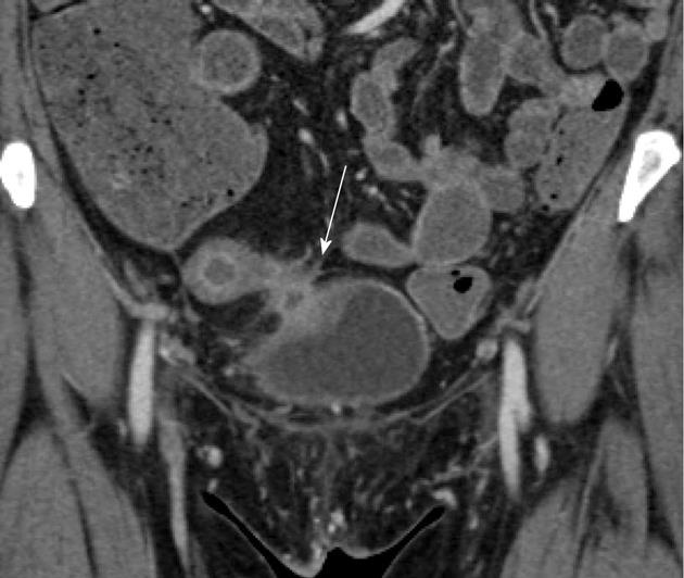
Figure 7 Enterovesicular fistula.
Fifty-two-year-old female with Crohn’s disease. Coronal contrast-enhanced computed tomography image demonstrates a markedly thickened, hyperemic loop of bowel in the pelvis, in keeping with acute Crohn’s related inflammation. The bowel loop directly abuts the bladder, which is focally thickened (arrow) at the site of contact, although no gas was identified in the bladder. The patient was ultimately proven to have an enterovesicular fistula.
- Citation: Raman SP, Horton KM, Fishman EK. Computed tomography of Crohn’s disease: The role of three dimensional technique. World J Radiol 2013; 5(5): 193-201
- URL: https://www.wjgnet.com/1949-8470/full/v5/i5/193.htm
- DOI: https://dx.doi.org/10.4329/wjr.v5.i5.193


