Copyright
©2013 Baishideng Publishing Group Co.
World J Radiol. May 28, 2013; 5(5): 193-201
Published online May 28, 2013. doi: 10.4329/wjr.v5.i5.193
Published online May 28, 2013. doi: 10.4329/wjr.v5.i5.193
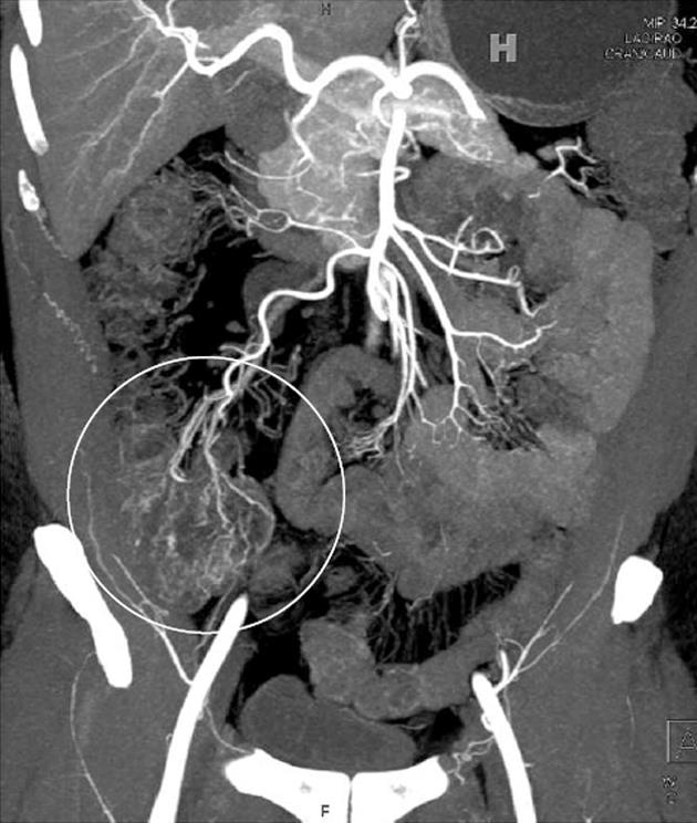
Figure 4 Identification of early Crohn’s disease using maximum intensity projection images.
Forty-seven-year-old male with abdominal pain. While no significant abnormality was appreciated on the axial source images or multiplanar reformats, coronal maximum intensity projection images raised the possibility of mild mesenteric hyperemia and vasa recta engorgement (circle) adjacent to the cecum. The patient underwent colonoscopy, and was found to have Crohn’s colitis.
- Citation: Raman SP, Horton KM, Fishman EK. Computed tomography of Crohn’s disease: The role of three dimensional technique. World J Radiol 2013; 5(5): 193-201
- URL: https://www.wjgnet.com/1949-8470/full/v5/i5/193.htm
- DOI: https://dx.doi.org/10.4329/wjr.v5.i5.193


